
The main achievements of the Institute
A smart nanomaterial capable of expanding the therapeutic window of radiation therapy without increasing the dose of radiation
In this study, stable nanocomposites based on the nanoparticles of cerium dioxide (CeO2) encapsulated in a PMAO–Jeffamine brush copolymer were obtained and experimentally characterized. These nanocomposites provide long-lasting and selective redox-activity in a cellular environment, as shown in the figure. Polymer encapsulation does not shield the catalytically active surface of CeO2 but preserves the ability of nanoparticles to undergo reversible Ce3+/Ce4+ transitions and autocatalytic interaction with reactive oxygen species, including after exposure to ionizing radiation.
The study demonstrates that the same nanomaterial exhibits opposite biological effects: radioprotective (antioxidant) in normal fibroblasts and pro-oxidant in breast tumor cells, as confirmed by a combination of methods (MTT, ROS analysis, mitochondrial potential, RT-PCR). The significance of these results lies in establishing a scientific foundation for the development of selective nanomaterials capable of expanding the therapeutic window of radiotherapy without increasing the dose of radiation. Thus, a smart nanomaterial has been developed that protects healthy cells while simultaneously enhancing damage to tumor cells.
Potential areas of application: radiation therapy for malignant tumors, development of theranostic nanoplatforms, radioprotectors for normal tissues, and biomedical nanocatalysts with controllable redox activity.
Technology readiness level: 3
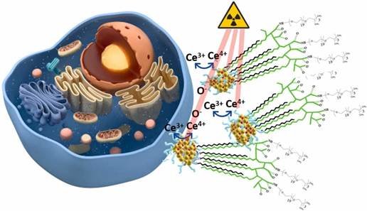
Schematic representation of a nanocomposite based on cerium oxide and PMAO–Jeffamine.
Organizations: Institute of Theoretical and Experimental Biophysics of the Russian Academy of Sciences, Saratov State University, National State Nuclear University MEPhI, Moscow Institute of Physics and Technology
Authors: Goryacheva О.А., Strokin P.D., Burov А.М., Presnyakov K.Y., Prikhozhdenko Е.S., Anikina V.А., Zamyatina Е.А., Romanov R.I., Chukavin N.N., Markeev А.М., Popova N.R.
Publication: Goryacheva O.A., Strokin P.D., Burov A.M., et al. Cerium Dioxide Nanoparticles in PMAO-Jeffamine Brush Copolymer for Sustained Redox Activity in Cellular Environments. Colloids and Surfaces A: Physicochemical and Engineering Aspects. 2026; 728:138545.
A new, highly effective biomimetic approach has been developed for creating materials with a considerable osteogenic, angiogenic, and hematopoiesis-inducing potential
An innovative method for producing bone-replacing materials has been developed, based on biomimetic deposition (remineralization) onto the surface of demineralized bone-collagen matrices with an intact natural ultrastructure (DBM) of low-temperature hydroxyapatite precursors (DBM+CaP) in the presence of serum albumin (DBM+CaP+BSA). Ectopic (subcutaneous) implantation of the materials to experimental animals was used to assess their true osteogenic potential. This approach yielded a remarkable and unprecedented result, demonstrating the pronounced angiogenic and osteogenic properties of the obtained composite materials. These properties consisted not only in the accelerated ectopic formation and mineralization of full-fledged new bone tissue and the formation of a mature vascular network (Fig. c, f, g), but also in the de novo formation of fully functional bone marrow in the intertrabecular space (Fig. a, b, d, e) with hematopoietic (blood-forming) activity. The proposed approach can be successfully used in the production of bioactive materials for spinal surgery or the surgical correction of large bone defects, since such materials can simultaneously serve as both an effective osteogenic scaffold and a source of progenitor cells.
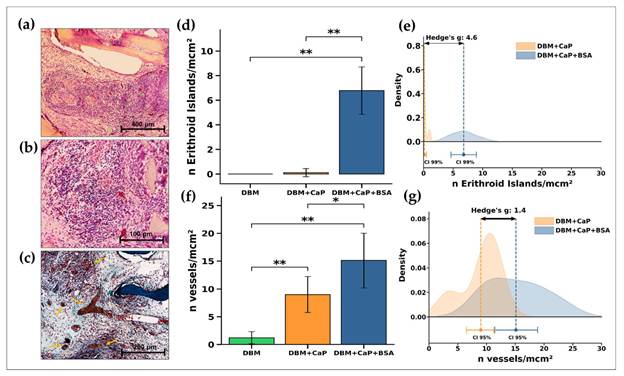
Histological and quantitative analysis of samples from the DBM + CaP + BSA group. (a-c) H&E and Lillie’s trichrome staining; (d) comparative analysis of the number of erythroid islands and (e) the corresponding Hedges’ g; (f) comparative analysis of newly formed blood vessels and (g) the corresponding Hedges’ g. A Hedges’ g value ≥ 0.80 indicates a pronounced effect. * – p < 0.05. ** – p < 0.01. CI — confidence interval.
Organizations: Institute of Theoretical and Experimental Biophysics of the Russian Academy of Sciences, in collaboration with the A.A. Baikov Institute of Metallurgy and Materials Science of the Russian Academy of Sciences.
Supervisors: I.S. Fadeeva, Leading Researcher and Head of the Laboratory at ITEB RAS, Ph.D in Biological Sciences; A.Yu. Teterina, Researcher at IMET RAS, Ph.D in Technical Sciences.
Authors: Minaychev V.V., Teterina A.Yu., Smirnova P.V., Menshikh K.А., Senotov А.S., Kobyakova М.I., Smirnov I.V., Pyatina K.V., Krasnov K.S., Fadeev R.S., Komlev V.S., Fadeeva I.S.
Minaychev V.V., Teterina A.Yu., Smirnova P.V., Menshikh K.A., Senotov A.S., Kobyakova M.I., Smirnov I.V., Pyatina K.V., Krasnov K.S., Fadeev R.S., Komlev V.S., Fadeeva I.S. Composite Remineralization of Bone-Collagen Matrices by Low-Temperature Ceramics and Serum Albumin: A New Approach to the Creation of Highly Effective Osteoplastic Materials. J. Funct. Biomater. 2024, 15, 27.
A hypothesis has been proposed and confirmed regarding the existence of two functionally distinct subpopulations of cancer lipid droplets. One of them operates intracellularly and forms during the cell division stage, while the other is released into the extracellular microenvironments from the surface of the cancer cell, thereby locally increasing the levels of extracellular lipids capable of suppressing the anti-tumor immune response. By utilizing this dual defense mechanism, aggressive cancer cells gain additional advantages for their survival and growth. The ability of cancer cells to serve as a source of both intracellular and extracellular lipids has made it possible to create a metabolic model illustrating the dual advantages gained by cancer cells (but not immune cells), as well as to identify potential targets for correcting this pathological condition (ITEB RAS, P.M. Schwartsburd, Doctor of Biological Sciences).
The results are published in Cancer communication: Polina Schwartsburd. Lipid droplets: could they be involved in cancer growth and cancer–microenvironment communications? Cancer Communications.2022;42:83–87. https://doi.org/10.1002/cac2.12257

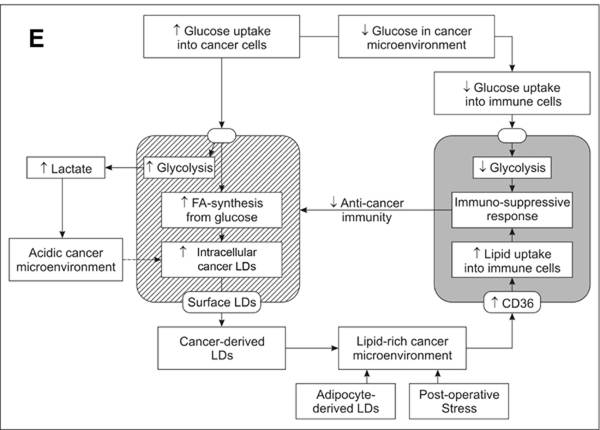
A model of metabolic interactions between cancer cells, their microenvironment, and immune cells. Cancer lipid droplets contribute to elevated lipid levels in cancer microenvironment, thereby promoting a reduction in the anti-tumor immune response.
It has been established that the condensation of flavonoids with toxic carbonyl compounds results in the formation of products exhibiting high antioxidant activity. New effective 3-aminopyridone-based luminophores have been discovered that can be used to develop sensitive methods for detecting hydrogen peroxide and to modify ELISA techniques (ITEB RAS, Shatalin Y.V., Ph.D in Biological Sciences).
The results are published in Antioxidants и Dyes and Pigments:
1. Shubina V.S., Kozina V.I., Shatalin Y.V. Comparison of Antioxidant Properties of a Conjugate of Taxifolin with Glyoxylic Acid and Selected Flavonoids. Antioxidants, 2021, 10, 1262. 10.3390/antiox10081262.
2. Shatsauskas A., Shatalin Y., Shubina V., Zablodtskii Y., Chernenko S., Samsonenko A., Kostyuchenko A., Fisyuk A. Synthesis and application of new 3-amino-2-pyridone based luminescent dyes for ELISA. Dyes and Pigments. 2021; 187:109072. 10.1016/j.dyepig.2020.109072.
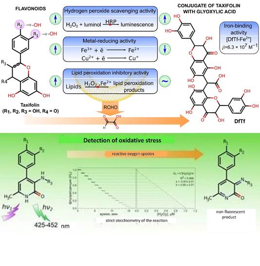
Relationship between the structure and antioxidant activity of the polyphenols under study. Structure of a new class of sensitive luminophores.
New ceramic biomaterials based on nanosized iron-substituted hydroxyapatites and biphasic ceramics containing α- and β-modifications of tricalcium phosphate (TCP) have been developed and studied, along with manganese-doped TCP-based bioactive ceramic coatings deposited onto a titanium surface by arc plasma spraying. Good adhesion, spreading, and high proliferative activity of human dental pulp stem cells were demonstrated on the surface of metal-substituted hydroxyapatites. A reduction in the proliferative activity of dental pulp cells on the surface of biphasic α, β-tricalcium phosphate ceramics was observed, attributed to changes in surface morphology (specifically, the formation of a lamellar crystalline layer). The developed biomaterials and coatings show promise for enhancing the biocompatibility and integration characteristics of bone implants in orthopedics and dentistry. (ITEB RAS, Davydova G.A., Selezneva I.I.)
1. Fadeeva I.V., Selezneva I.I, Davydova G.A., Fomin A., Gafurov M., Barinov S.M., Poltavtsev A.V., Davydova I.B., Zaraisky E.I., Poltavtseva R.A. Nanosized iron-substituted hydroxyapatites // IOP Conference Series Materials Science and Engineering 747, 2020, 012066.
2. T.V. Safronova, I.I. Selezneva, S.A. Tikhonova, A.S. Kiselev, G.A. Davydova, T.B. Shatalova, D.S. Larionov, J.V. Rau. Biocompatibility of biphasic α,β-tricalcium phosphate ceramics in vitro// Bioactive Materials, Volume 5, Issue 2, June 2020, Pages 423-427.
3. I. V. Fadeeva, V. I. Kalita, D. I. Komlev, A. A. Radiuk, A. S. Fomin, G.A. Davidova, N. K. Fursova, F. F. Murzakhanov, M. R. Gafurov, M. Fosca, I. V. Antoniac, S.M. Barinov, J. V. Rau. In Vitro Properties of Manganese-Substituted Tricalcium Phosphate Coatings for Titanium Biomedical Implants Deposited by Arc Plasma// Materials (Basel), 2020, Oct 3; 13(19):4411.
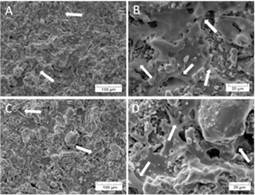
Images of dental pulp stromal stem cells cultivated for 2 days on the surface of the following materials: TCP (A, B) and Mn-TCP (C, D). Pseudopodia are indicated by arrows.
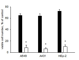
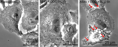
Research is underway to develop combination anti-tumor therapies using a combination of known drugs, specifically vitamin B12b together with diethyldithiocarbamate (DDC), a compound related to the anti-alcohol drug Diacarb. It has been found that vitamin B12b and DDC, when added at non-toxic concentrations, induce tumor cell death—a finding that could serve as the basis for developing a drug for combination anti-tumor therapy. Both of these drugs have long been used in medicine and have minimal undesirable side effects, which facilitates further research into the effects of their combination. (Institute of Theoretical and Experimental Biophysics of the Russian Academy of Sciences (ITEB RAS), Solovieva М.Е., Shatalin Y.V., Solovyev V.V., Sazonov А.V., Kutyshenko V.P., Akatov V.S.)
a
b
Potentiation of the toxic effect of diethyldithiocarbamate by vitamin B12b. Black bars indicate tumor cell survival after a 48-h incubation with 1 мМ DDC; white bars indicate a decrease in the number of living cells upon the addition of В12b together with DDC (а). Vacuolization of А549 cells (arrows indicated vacuoles) following a 5-h incubation with DDC+B12b, preceding their death (b).
The results are published in Redox Biology: Solovieva ME, Shatalin YV, Solovyev VV, Sazonov AV, Kutyshenko VP, Akatov VS. Hydroxycobalamin catalyzes the oxidation of diethyldithiocarbamate and increases its cytotoxicity independently of copper ions. Redox Biology, 2019, 20, pp. 28-37. IF = 7.793
The study demonstrates the involvement of the NF-κB transcription factor signaling cascades and the mTOR protein signaling complex 1 (TORC1) in the acquisition of resistance by leukemic cells to receptor-mediated apoptosis and to drugs in multicellular structures.
It has been shown that inhibition of only a group of anti-apoptotic proteins from the Bcl-2 family—Bcl-2, Bcl-XL, Bcl-w, Bfl-1, and Mcl-1—significantly suppresses the resistance of leukemia cells to TRAIL-mediated apoptosis and apoptosis induced by topoisomerase inhibitors in multicellular structures.
Specific differences in the proliferation rates of human atrial and ventricular fibroblasts (HVFs) are demonstrated, along with simvastatin-mediated inhibition of HVF proliferation via the HMG-CoA reductase pathway, which is reversibly abolished by the addition of mevalonic acid.
(ITEB RAS, Professor V.S. Akatov, Doctor of Physical and Mathematical Sciences)
Despite many years of effort, no methods have yet been developed to prevent or slow the progression of acquired epilepsy in at-risk groups, indicating an insufficient understanding of the mechanisms of epileptogenesis. Acquired epilepsy can result from a range of pathogenic insults to the brain (strokes, traumas, etc.) or from neurological diseases with a wide variety of etiologies. This implies that, despite their diversity, all these initiating pathologies affect the same fundamental brain functions underlying network excitability. Identifying these mechanisms and their therapeutic accessibility is essential for developing effective strategies for the prevention and treatment of epileptogenesis. Based on extensive experimental evidence, we propose a hypothesis regarding the key role of NADPH oxidase-mediated oxidative stress and glucose hypometabolism in neural tissue in the initiation of epileptogenesis and seizure activity; furthermore, we offer a new perspective on the mechanisms underlying acquired epilepsy and identify targets for potential therapeutic interventions. (ITEB RAS, I.Yu. Popova, Ph.D in Biology).
The results are published in Trends in Pharmacological Sciences: Yuri Zilberter, IrinaPopova, MishaZilberter. Unifying mechanism behind the onset of acquired epilepsy. Trends Pharmacol Sci, 2021 Dec 6; S0165-6147(21)00226-1.
The article was selected for the journal’s cover.
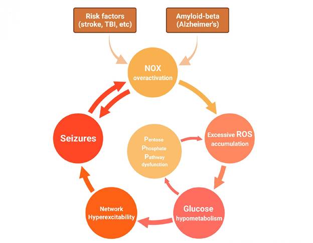
The diagram illustrates the relationship between oxidative stress, glucose hypometabolism, and seizures in acquired epilepsy.
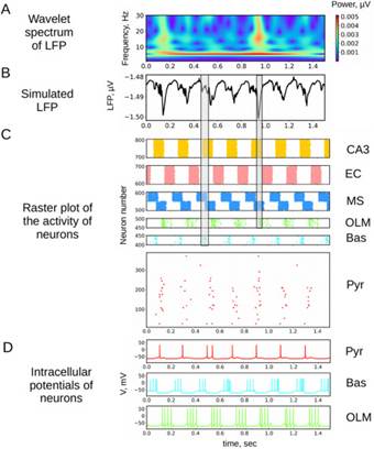
The hippocampus plays a critical role in attention and memory processes. When the brain processes new information, a theta rhythm—potential oscillations at a frequency from 4 to 12 Hz—is observed in the hippocampus. A mathematical model of a neural network representing one of the hippocampal regions (CA1 field) in the theta rhythm generation mode has been proposed. The model accounts for the main neuronal populations of the hippocampus and the arrival of signals via external inputs. In computational experiments, the model reproduces the synchronization parameters of neuronal populations and the main electrophysiological characteristics of the theta rhythm described in the experimental literature. The results obtained allow us to draw conclusions about the contribution of external inputs and CA1 neurons to the generation of potential oscillations. In particular, it was shown that an excitatory input to the CA1 field from another region of the hippocampus, the CA3 field, plays the most important role in the generation of the theta rhythm. It should be noted that the proposed model not only reproduces the characteristics of the theta rhythm but also identifies which hippocampal neuronal groups are involved in information transfer and the timing of such transfer.
The results are published in: Mysin I.E., Kitchigina V.F., Kazanovich Y.B. Phase relations of theta oscillations in a computer model of the hippocampal CA1 field: Key role of Schaffer collaterals. Neural Network. 2019, 116, 119-138. https://www.sciencedirect.com/science/article/pii/S0893608019301029?via%3Dihub
This study continues a series of investigations into the mechanisms underlying the development of epilepsy, one of the most common and heterogeneous neurological disorders. Reduced cerebral glucose uptake is an early and widespread symptom of all major neurodegenerative diseases, including acquired epilepsy. However, the mechanisms driving this inhibition of glucose metabolism remain unclear. In this study, we demonstrated that seizure activity leads to oxidative stress in nervous tissue, followed by rapid and sustained inhibition of glucose uptake. Thus, seizures promote glucose hypometabolism in the brain, creating a positive feedback loop. The study demonstrates that normalizing glucose metabolism in the brain—for instance, using exogenous energy substrates—is essential to halt this pathological cycle. This research may serve as a foundation for effective therapies for seizure disorders. The work was conducted as part of an international collaboration with Unit 751 of the French National Institute of Health and Medical Research (INSERM UMR1106, Institut de Neurosciences des Systèmes, Aix-Marseille University, Marseille, France). (ITEB RAS, A.E. Malkov, Ph.D in Biology).
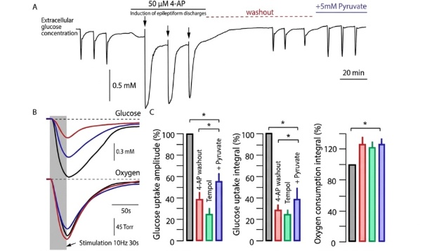
Reduction in glucose uptake following the induction of seizure discharges in surviving brain slices from experimental animals.
А) Extracellular glucose concentration measured using a polarographic sensor in the CA1 field of the hippocampus. After recording control responses to electrical stimulation of Schaffer collaterals (10 Hz, 30 s, 120 μA), 4-AP was applied and seizure discharges were induced. After a prolonged washout period of at least 40 min, responses to control stimulation were recorded. Glucose uptake was significantly reduced. After adding pyruvate (5 mM) to the medium, glucose uptake returned to control levels.
(B; C) Changes in extracellular glucose concentration and oxygen partial pressure in response to stimulation. Control responses are shown in black; responses to stimulation after 4-aminopyridine washout are shown in red; changes following the addition of pyruvate are shown in blue. Unlike pyruvate, the antioxidant Tempol (indicated in green) did not have a restorative effect on glucose uptake.
The results were published in Neurobiology of Disease: Malkov A, Ivanov AI, Buldakova S, Waseem T, Popova I, Zilberter M, Zilberter Y. Seizure-induced reduction in glucose utilization promotes brain hypometabolism during epileptogenesis. Neurobiol Dis. 2018 Aug;116:28-38. doi: 10.1016/j.nbd.2018.04.016.
Epilepsy is one of the most common and heterogeneous neurological disorders. Metabolic dysfunctions are a characteristic feature of various types of epilepsy in humans and, moreover, are used to localize the seizure focus in patients. This study tested the hypothesis that impaired neuronal energy homeostasis is the root cause of seizure activity in epilepsy and Alzheimer’s disease, and that supplying the brain with an alternative energy source—other than glucose—could be an effective therapeutic strategy for seizure disorders. In three different models of chronic acquired epilepsy (focal, generalized, and associated with Alzheimer’s disease), it was demonstrated that chronic oral administration of pyruvate, an anaplerotic mitochondrial substrate, exerts a potent antiepileptic effect by normalizing brain electrical activity and metabolic parameters. These findings may serve as a foundation for effective therapies of seizure disorders. The work was conducted as part of an international collaboration with Unit 751 of the French National Institute of Health and Medical Research (INSERM UMR1106, Institut de Neurosciences des Systèmes, Aix-Marseille University, Marseille, France). (ITEB RAS, I.Yu. Popova, Ph.D in Biology).
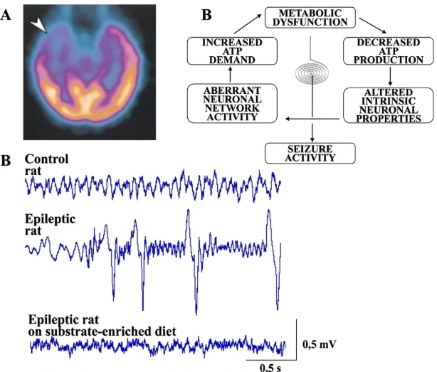
А. Glucose hypometabolism in the epileptic brain (PET method, from Pan JW et al., 2000). B. A schematic representation of the hypothesis regarding the development of seizure activity associated with metabolic dysfunction, based on the principle of positive feedback. C. Potent antiepileptic effect of metabolic therapy. Records of hippocampal electrical activity in three groups of experimental animals are shown.
A study of the neuroprotective effect of the anti-inflammatory cytokine IL-10 on hypoxia-induced neuronal damage showed that, unlike immune system cells, IL-10 exerts its protective effect in neurons without the involvement of the PI3K/Akt intracellular signaling pathway. This confirms the non-canonical action of this interleukin on neurons, highlighting the need to identify other intracellular mediators of IL-10’s effects.
In the context of kainate-induced excitotoxic damage to hippocampal neurons, a reciprocal relationship has been demonstrated between the activity of metabotropic glutamate receptors (mGluRs) and their expression levels. This allows for predicting the efficacy of ligands targeting pre- and postsynaptic mGluRs as neuroprotective agents under conditions of glutamate excitotoxicity.
(ITEB RAS, V.I. Arkhipov, Doctor of Biological Sciences)
It has been shown that between 2% and 13% of the human population are “X-men” who possess an unusual configuration of receptor systems that allow them to construct a virtual world of new images within their brains. This phenomenon is known as synesthesia.
As early as the beginning of the 20th century, academician P. P. Lazarev intuitively proposed the idea that such a synthesis was possible, having examined just one specific form of synesthesia. At the time, this discovery was not understood and was dismissed as false. Half a century later, the existence of synesthesia was proven, and today, interest in its study has grown significantly.
It has been shown that synesthesia significantly reduces the time required to memorize and analyze large amounts of information.
It is not a pathology and may have a genetic basis or be acquired through intensive, purposeful engagement in creative processes (such as those of artists, scientists, architects, etc.)
The emergence of synesthesia is associated with a reorganization of cortical brain structures stimulated by hippocampal waves. This leads to the formation—via combinatorial strengthening of connections in the cerebral cortex—of links between the areas representing various receptor systems (see figure). These reorganizations in brain connections allow for the transformation of the multiparametric phenomena observed in the external world into generalized virtual images—forms that differ from one another in their topology or color. (Institute of Theoretical and Experimental Biophysics of the Russian Academy of Sciences (ITEB RAS), G.R. Ivanitskii, Corresponding Member of the Russian Academy of Sciences.)
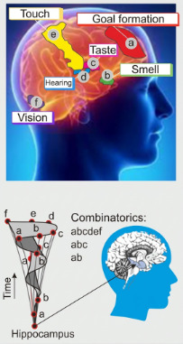
Reorganization of cortical brain structures stimulated by hippocampal waves, leading to the formation of connections in the cerebral cortex between areas representing different receptor systems through combinatorial reinforcement.
The results are published in Uspekhi Fizicheskikh Nauk (Advances in Physical Sciences):
Ivanitskii G.R. Ludi X, obladayushchiye neobychnym vzaimodeistviyem retseptornykh system, construiruyut vnutri sebya mir novykh obrazov (k 140-letiyu akademika P.P. Lazareva [X-men: humans with an unusual interaction between receptor systems who construct a world of new images within themselves (on the 140th anniversary of the birth of academician P.P. Lazarev)] / Uspekhi Fizicheskikh Nauk 189(7) p.759-784 (2019)
Characteristic biological constants have been identified that determine the limits of temperature adaptation for most organisms with a nervous system, including invertebrates, hibernators, and even humans. These constants (6.5°C, 27°C, and 43°C) make it possible, for instance, to induce and maintain hypometabolic states following major surgical procedures, as well as during long-duration space flights. The study identified the following general biological patterns:
• The phase transition temperature range for the lipid-protein composition of the presynaptic membrane at [Ca+2] > 10⁻¹ mM extends from 6.5°C to 27°C.
• The phase transition temperature range for the lipid-protein composition of the presynaptic membrane at [Ca²⁺] < 10⁻⁴ mM extends from 27°C to 43°C.
• A 1000-fold increase in Ca²⁺ concentration corresponds to a 15°C decrease in the phase transition temperature of the most high-melting lipid-protein compositions of the presynaptic membrane and a 20°C decrease in the most low-melting compositions.
• The ratio of the limiting temperature ranges TTP-(TC)min/(TH)max-TTP
= 1.3 is determined by the phase transition temperatures of the lipid-protein compositions of synaptic membranes during excitation and at rest. (ITEB RAS, Aslanidi K.B., Doctor of Biological Sciences)
The results are published in Cell & Bioscience: Aslanidi K.B., Kharakoz D.P. Limits of temperature adaptation and thermopreferendum. Cell Biosci. 2021 Apr 6;11(1):69. doi: 10.1186/s13578-021-00574-9.
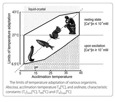
Temperature adaptation of various organisms. Аbscissa, acclimation temperature ТА[0C], and ordinate, characteristic biological constants (ТС)min [0C], TPR[0C] и (ТА)max[0C]
It has been shown that low-amplitude transcranial electrical stimulation affects cognitive function: specifically, the solving of logical, geometric, and arithmetic problems. The outcome depends on the polarity of the stimulus: anodal stimulation leads to improved cognitive performance, whereas with cathodal stimulation, the effect is barely discernible. Anodal stimulation enhances cortical electrical activity, contributes to greater cognitive efficiency, and optimizes the autonomic mobilization of resources during cognitive tasks. Cathodal stimulation affects the spectral power of the EEG depending on the individual balance of excitation and inhibition in the brain and increases the influence of the parasympathetic branch of the autonomic nervous system. Thus, low-amplitude transcranial electrical stimulation, as a promising non-invasive method, can be used to improve cognitive function. (ITEB RAS, Aliev R.R., Doctor of Physical and Mathematical Sciences).
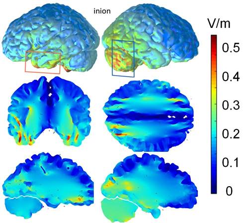
Distribution of the electric field generated by the active (red) and reference (blue) electrode placements during a low-amplitude transcranial stimulation procedure
The results are published in: Brain Stimulation, impact factor 6.078.
Ashikhmin A. V., Shishelova A. Y., Aliev R. R. tDCS provokes sustainable changes in EEG and reorganizes autonomic modulation of heart rate //Brain Stimulation: Basic, Translational, and Clinical Research in Neuromodulation. 2017. 10 (№. 2). 484-486.
Interfacial membranes between phases consist of a network of three-dimensional protein cells filled with lipids mixed with bound water. Both in model and in vivo experiments at the molecular and cellular levels, agents have been identified that can increase or decrease the stability of phase boundaries. In particular, it has been shown that taxifolin and kaempferol act as stabilizing agents, accelerating the formation of fibrils in the network, whereas quercetin and myricetin inhibit protein aggregation and hinder fibril formation. The kinetics of phase boundary changes can be controlled not only by altering the chemical composition of the agents but also through physical interventions (electromagnetic irradiation, temperature, pressure). A kinetic model has been developed that encompasses the main molecular events in networks, including protein-protein interactions, the activation and deactivation of GTPases, phosphorylation and dephosphorylation, as well as feedback regulation. Methods for raising or lowering the barriers that maintain the stability of membrane boundaries have practical significance as tools for modern medicine, cell and tissue engineering, as well as in artificial insemination for the conservation of endangered species (as demonstrated using amphibians as an example).
(ITEB RAS Ivanitskii G.R., Corresponding Member of the Russian Academy of Sciences)
In 2016, based on data on the thermodynamic parameters of proteins, hydration parameters at a temperature of 298 K were used to estimate the numerical values of the enthalpy, free energy, and entropy of these proteins in the gas phase. Statistical studies were conducted on the dependence of the frequency of bond breaks on the DNA nucleotide sequence, which allows for the assessment of errors in experimental procedures used to determine the primary structures of DNA. Cell growth on various polymers and the detachment of cell layers from these surfaces were investigated. Furthermore, experiments were conducted involving long-term synchronous monitoring of minute-by-minute heart rate values and blood biochemical parameters; the findings highlighted the significant role of a number of biochemical factors in the body’s adaptation to variations in the geomagnetic field. A comparative study of the "all-combinations" method, the minimal-covering method, and the histogram method was conducted. Intraday periods in time series of alpha-decay rate fluctuations were investigated, and their connection to the long-wavelength part of the Earth’s natural oscillation spectrum was demonstrated. New aspects of solving the problem of auto-wave front refraction at the interface between two homogeneous regions of an active medium were described. Manifestations of a universal system of tripling periods were found across a wide range of natural and technical systems. The nature of genetic variability in P. pica from Europe, the Far East, Japan, Korea, and Taiwan was described. An analysis of the bioacoustic parameters of P. pica signals was performed in comparison with genetic results. Mechanisms of isolation have been identified in the closely related species Acrocephalus dumetorum and Acrocephalus orinus.
Based on the database created and the rules discovered for how proteins recognize specific DNA segments, it is possible to design therapeutic agents for the treatment of genetic diseases.
As part of the IDEA-2016 competition, a Russian manufacturer of medical devices was provided with effective frequency values for magnetotherapy and UHF therapy, which were obtained through the study of a universal system of tripling periods.
(ITEB RAS, Polozov R.V., Doctor of Physical and Mathematical Sciences)
Studies on the isoform composition and functional properties of muscle proteins—conducted under normal conditions as well as during adaptation and pathological processes using a suite of methods (including fluorescence analysis, spectrophotometry, circular dichroism, dynamic light scattering, X-ray diffraction, and confocal, electron, and atomic force microscopy)—have demonstrated that the giant protein titin in striated and smooth muscles forms amyloid aggregates in vitro. A toxic effect of titin amyloids on smooth muscle cell cultures was observed. The results suggest that titin may play a role in the formation of cytotoxic aggregates in vivo during the development of amyloidoses.
(ITEB RAS, Vikhlyantsev I. M., Doctor of Biological Sciences)
A new experimental and theoretically grounded method for the in vitro evaluation of osteoplastic materials has been developed, successfully replacing their implantation in laboratory animals.
It has been demonstrated for the first time that the use of a simple mixture of DMEM medium and fetal bovine serum, incubated in a custom-designed perfusion-diffusion bioreactor, enables the in vitro reproduction of the physicochemical changes that occur in calcium-phosphate materials upon their implantation into the body (Fig.). This bioreactor overcomes mass-transfer limitations at the material-liquid interface and simulates natural blood or interstitial fluid flow, unlike the standard SBF model. Within the body, a complex bilateral material-medium interaction takes place, manifested by the release of calcium and phosphate ions into body fluids, the adsorption of amino acids and proteins by the surface of the material, and phase transformations (e.g., from dicalcium phosphate dihydrate (DCPD) to octacalcium phosphate (OCP)), which significantly influences bone defect regeneration. In this study, these processes were described mathematically in terms of chemical kinetics, which allows for a numerical assessment of the influence of each process on in vivo osseointegration of bone implants. Thus, the developed method helps to reduce the number of animal experiments, complies with the international 3R principle (Replacement, Reduction, Refinement), and may serve as a step toward personalized medicine. This approach integrates biochemistry and patient medical history with numerical modeling and/or experimental techniques to select the optimal drug therapy or implants.
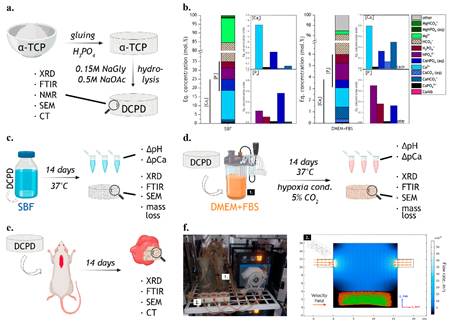
Experimental setup: (a) synthesis method; (b) characteristics of the media used: distribution of ionic forms in SBF and in 90% DMEM + 10% FBS; (c) schematic of Model I and analysis methods used; (d) schematic of Model II and analysis methods used; (e) schematic of the in vivo experiment and analysis methods used; (f) appearance of the bioreactor and velocity distribution in the culture chamber.
Organizations: Institute of Theoretical and Experimental Biophysics of the Russian Academy of Sciences, in collaboration with the A.A. Baikov Institute of Metallurgy and Materials Science of the Russian Academy of Sciences.
Supervisors: A.Yu. Teterina, Researcher at IMET RAS, Ph.D in Technical Sciences; I.S. Fadeeva, Leading Researcher and Head of the Laboratory at ITEB RAS, Ph.D in Biology.
Authors: Shlykov M.A., Smirnova P.V., Senotov A.S., Teterina A.Y., Minaychev V.V., Smirnov I.V., Novikov R.A., Marchenko E.I., Salynkin P.S., Komlev V.S., Fadeev R.S., Fadeeva I.S.
Shlykov M.A., Smirnova P.V., Senotov A.S., Teterina A.Y., Minaychev V.V., Smirnov I.V., Novikov R.A., Marchenko E.I., Salynkin P.S., Komlev V.S., Fadeeva I.S. Comparative Evaluation of Mathematical Model and In Vivo Study of Calcium Phosphate Bone Grafts. J. Funct. Biomater. 2024, 15(12), 368.
Among various drug development strategies, repurposing existing drugs for new diseases is a promising approach, as these drugs have already undergone preclinical and clinical trials and are well understood in terms of safety and side effects.
This study tested the drug Tameron (monosodium α-luminol or the sodium salt of 5-amino-2,3-dihydro-1,4-phthalazinedione)—a new, unpatented drug also known as Galavit or MP1032. Given the positive results achieved with Tameron as an antiviral, immunomodulatory, and anti-inflammatory agent with an antioxidant mechanism of action, it was decided to further investigate its role as a potential antioxidant and radioprotective agent.
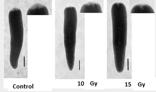
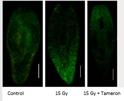
Planarian flatworms—invertebrates with an extraordinary capacity for regeneration due to their bodies being rich in stem cells known as neoblasts (comprising up to 30% of their cells)—were selected as the experimental model. The neoblasts endow the planarian body with virtually unlimited regenerative potential, enabling the animals to replace missing body parts or even regenerate an entire worm from a small body fragment. Exposing planarians to sublethal X-ray doses, preserving a part of neoblasts with a potential for further regeneration, is the basis of our experimental model (Fig. 1).
In the course of the study, Tameron proved to be a powerful new radioprotector and antioxidant that significantly increased the rate of regeneration in planarians damaged by X-ray- or menadione-induced oxidative stress and protected the mitotic activity of neoblasts (Fig. 2). It also modulated the expression patterns of neoblast marker genes and NRF-2-regulated oxidative stress marker genes.
The results are published in Antioxidants (Basel): Tsarkova E., Filippova K., Afanasyeva V., Ermakova O., Kolotova A., Blagodatski A., Ermakov A. A Study on the Planarian Model Confirms the Antioxidant Properties of Tameron against X-ray- and Menadione-Induced Oxidative Stress. Antioxidants (Basel). 2023 Apr 18;12(4):953. doi: 10.3390/antiox12040953. IF 7
|
|
|
|
Fig.1. Representative photographs of regenerating planarians used to analyze blastema growth rate. The analysis was performed using computer morphometry. The images show planarians on the third day after decapitation; control planarians and those exposed to X-ray radiation at doses of 10 and 15 Gy. Space bar: 1 mm. |
Fig.2. Mitotic cells in planarian bodies (immunocytochemical staining) 7 days after irradiation at a dose of 15 Gy. Space bar is 1 mm. |
A system for the continuous detection of various types of pathogens with high speed and sensitivity
A system has been developed that enables the real-time detection of pathogen biomarkers in a sample stream. High performance of the method is ensured by the use of a unique active microarray-based immunoassay technology employing magnetic labels, developed at the ITEB RAS and validated by publications in high-impact journals. Its primary area of potential application is biosafety, specifically the prevention of bioterrorism and environmental monitoring. Key features of the system include: 1) ultra-high sensitivity, capable of detecting trace biomarker concentrations on the order of tens of thousands of molecules per milliliter of sample; 2) short analysis time, with a positive signal received within 10 minutes of the pathogen entering the system; 3) the ability to operate continuously for extended periods (at least 12 hours); and 4) the capability for full automation. The figure shows a photograph of the device.
Based on the combination of these characteristics, the system is unique and enables the detection of a wide range of bacterial pathogens. Furthermore, the method can be adapted to detect intact viral particles and specific nucleic acid fragments
Technology Readiness Level: 4
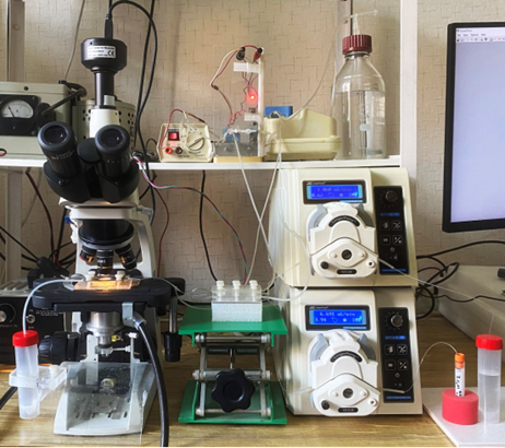
A system for the continuous detection of various types of pathogens.
Institute of Theoretical and Experimental Biophysics of the Russian Academy of Sciences
Authors: Shlyapnikov Y.M., Shlyapnikova E.A.
Shlyapnikov Y.M., Shlyapnikova E.A. Ultrasensitive Bead-Based Immunoassay for Real-time Continuous Sample Flow Analysis. Biosensors (Basel), 2025, 15(5), 316.
Cerium oxide nanoparticles modified with pyrroloquinoline quinone exhibit selective cytotoxicity upon X-ray irradiation
The study demonstrated that CeO₂@PQQ nanoparticles exert a selective cytotoxic effect on murine EMT6/P mammary adenocarcinoma cells when combined with X-ray irradiation, while protecting normal murine L929 fibroblast cells. In normal cells, the nanoparticles promoted the restoration of mitochondrial function and antioxidant levels, whereas in cancer cells, the nanoparticles contributed to a decrease in glutathione levels and mitochondrial membrane potential. As shown in the figure, CeO₂@PQQ nanoparticles increase the formation of micronuclei—a marker of DNA damage—in EMT6/P cancer cells following X-ray irradiation. Thus, it can be hypothesized that the nanoparticles may enhance DNA damage in cancer cells induced by ionizing radiation. The results of the study suggest that CeO₂@PQQ nanoparticles may play a key role in the development of more effective and safer methods for treating cancer due to the selective toxic effect of the nanoparticles on cancer cells and their protective effect on normal cells.
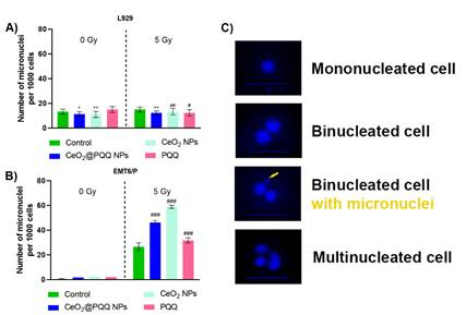
Number of micronuclei in L929 (A) and EMT6/P (B) cells after incubation with CeO2@PQQ, CeO2 NP, and PQQ at a concentration of 2 µM. Representative images of cell nuclei and micronuclei stained with DAPI are shown (the yellow arrow indicates a micronucleus) (C). Here, 0 Gy corresponds to the groups that were not irradiated, while 5 Gy corresponds to the groups exposed to X-ray irradiation at a dose of 5 Gy. Data are presented as M ± SD and were analyzed using Welch’s t-test (n = 5) (** p < 0.0051; *** p < 0.001 vs. the non-irradiated control; ### p < 0.001 vs. the irradiated control).
Organizations: Institute of Theoretical and Experimental Biophysics of the Russian Academy of Sciences (ITEB RAS) and Saratov National Research State University named after N.G. Chernyshevsky
Supervisor: Popova N.R., Senior Researcher and Head of Laboratory at the ITEB RAS, Ph.D in Biology.
Authors: Zamyatina Е.А., Goryacheva О.А., Popov А.L., Popova N.R.
Zamyatina, E.A.; Goryacheva, O.A.; Popov, A.L.; Popova, N.R. Novel Pyrroloquinoline Quinone-Modified Cerium Oxide Nanoparticles and Their Selective Cytotoxicity Under X-Ray Irradiation. Antioxidants 2024, 13, 1445.
Ultra-local thermodynamic control of electrophysiological processes in living cells: new fundamental possibilities in cell physiology
In the present study, a patch clamp experimental setup was developed for the first time that integrates a fully optical ultra-local temperature control in micro- and nano-volumes of a living cell. Optical temperature control was performed using synthetized graphitized nano-/micro-diamonds containing silicon vacancy color centers, which served as temperature sensors. The diamonds were mounted at the tip of a glass micropipette. The graphitic shell of the diamond allowed it to be heated via a tunable 640 nm laser, which simultaneously excited the silicon vacancy color centers.
The diamond’s temperature was determined by the shift of the zero-phonon line (at a frequency of 738 nm). The diamond was pre-calibrated for both heating and temperature. Thus, the nano-/micro-sized diamond functioned as a heater-thermometer at laser powers exceeding 1 mW—measuring its own temperature, which dropped sharply to the baseline solution temperature at micron-scale distances from the diamond in the aqueous medium. At low laser excitation power (less than 1 mW), the diamond did not heat up and remained capable of measuring the temperature of the surrounding environment. A schematic of the device and micrographs are shown in the figure.
This approach makes it possible, for the first time, to influence the electrophysiological processes in a living cell using ultra-local temperature profiles. This work is the first to include temperature in the privileged set of ultra-local electrochemical potential parameters, alongside the control of the cell membrane’s electrical potential and intracellular concentrations of calcium and other ions.
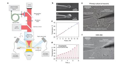
a) Schematic of the optical setup; b) a nanodiamond at the tip of a micropipette; с) calibration of nanodiamond heating based on laser power; d) protocol for applying thermal steps to the diamond; е-f) experimental configuration with living cells: a patch clamp pipette is on the left, a pipette with a nanodiamond is on the right.
Organizations: Prokhorov General Physics Institute of the Russian Academy of Sciences, Institute of Higher Nervous Activity and Neurophysiology of the Russian Academy of Sciences, Institute of Theoretical and Experimental Biophysics of the Russian Academy of Sciences.
Authors: Romshin А.М., Aseyev N.А., Idzhilova О.S., Koryagina А.А., Zeeb V.E., Vlasov I.I., Balaban P.М.
Romshin A., Aseyev N., Idzhilova O., Koryagina А., Zeeb V., Vlasov I., Balaban P. Rapid neurostimulation at the micron scale with an optically controlled thermal-capture technique. Biomater Sci, 2024 Nov 12
This work analyzes data demonstrating how unique fundamental properties of the protein bacteriorhodopsin can be used to create various types of practical devices—specifically, those designed for recording, processing, and storing optical information. It presents an overview of light-sensitive systems based on bacteriorhodopsin, summarizing current understanding of this protein, its properties, and its photocycle. The methods for modifying bacteriorhodopsin are discussed, including dehydration, the use of chemical additives, genetic alteration of its primary sequence, and the replacement of its natural chromophore with synthetic analogs. Such modifications can optimize the use of bacteriorhodopsin for the creation of light-sensitive recording media. Furthermore, the potential for using bacteriorhodopsin-based polymer films is examined, not only as photochromic materials for multiple recording, but also as a material for write-once recording and permanent memory. Although this type of memory and information processing is now considered exotic, the properties described in this work may serve as a foundation for developing unique light-sensitive hybrid technical devices that incorporate bacteriorhodopsin alongside other biological components.
The results are published in Journal of Photochemistry & Photobiology, C: Photochemistry Reviews: Druzhko Anna B. Some Aspects of Using the Fundamental Properties of Bacteriorhodopsin for Recording, Processing, and Storage of Optical Information. Journal of Photochemistry & Photobiology, C: Photochemistry Reviews, 56 (2023) 100620 https://www.sciencedirect.com/science/article/abs/pii/S1389556723000515?dgcid=rss_sd_all IF 13,6
|
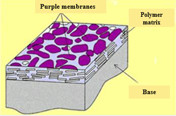
Fig. 1 Schematic representation of Biochrome film.

Fig. 2 Image obtained by projecting a negative stencil onto Biochrome film using yellow light.
Polyelectrolyte microcapsules (PMCs) are used in the development of targeted delivery systems, self-healing coatings, and diagnostic and theranostic systems. The protonation state of such complex systems can affect both the structure of the system itself and the substances encapsulated within it. A hypothesis was proposed and confirmed that the buffering properties of the microcapsules are determined by uncompensated segments of polyallylamine (PAA) in their composition. It was established that the buffer capacity of the capsules increases nonlinearly with the number of layers in the PMC shell. Furthermore, the buffer capacity of PMCs was found to depend on their ionic environment and incubation temperature. It was also found that PMCs disintegrate in a solution of sodium dodecyl sulfonate (SDS); this is likely due to electrostatic interaction between SDS molecules and the uncompensated PAA segments. These findings can be used to develop systems for targeted delivery and the sustained release of substances. Based on the data obtained, a diagnostic system has been developed for the rapid, cost-effective, and highly selective qualitative and semi-quantitative determination of SDS. (ITEB RAS, S.А. Tikhonenko, Ph.D in Biology)
The results are published in International Journal of Molecular Sciences и Scientific Reports:
· Alexey V. Dubrovskii et al. A Study of the Buffer Capacity of Polyelectrolyte Microcapsules Depending on Their Ionic Environment and Incubation Temperature. // Int. J. Mol. Sci. 2022, 23(12), 6608;
· Egor V. Musin et al. A Study of the Buffer Capacity of Polyelectrolyte Microcapsules Depending on Their Concentration and the Number of Layers of the Polyelectrolyte Shell // Int. J. Mol. Sci. 2022, 23(17), 9917;
· Kim, A.L., Musin et al. Qualitative and quantitative methods detection of SDS based on polyelectrolyte microcapsules.// Sci Rep 12, 232 (2022).
· Alexey V. Dubrovskii et al. Destruction of polyelectrolyte microcapsules and release of FITC-dextran from them by the influence of sodium dodecyl sulfonate // Sci Rep 12, 4032 (2022).
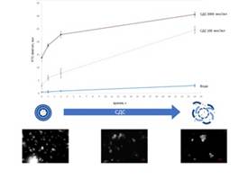
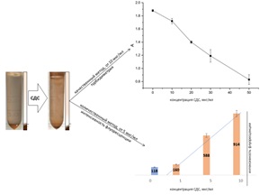
(I) Disruption of microcapsules using SDS; (II) Qualitative and quantitative analysis system for SDS based on microcapsules.
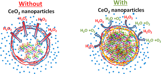
The development of effective systems for molecular diagnostics and targeted drug delivery is one of the most promising approaches to treating major diseases. In this study, we demonstrate the efficient encapsulation of luciferase in biodegradable polyelectrolyte microcapsules modified with therapeutically active cerium dioxide nanoparticles. It has been shown that modifying the microcapsules with citrate-stabilized cerium dioxide nanoparticles ensures that the encapsulated protein retains high activity when exposed to a damaging agent—hydrogen peroxide. This microcapsule structure not only ensures the preservation of the encapsulated substance under harsh environmental conditions but also enables its sustained release. The incorporation of redox-active nanoparticles into microcapsule structures will enable the development of new diagnostic and therapeutic agents for the effective diagnosis and treatment of a wide range of major diseases.
(ITEB RAS, Popov A.L., Ph.D in Biology)
The results are published in ACS Applied Materials & Interfaces serves:
Popov A., Popova, N., Gould D., Shcherbakov A.; Sukhorukov G.; Ivanov V. Ceria nanoparticles-decorated microcapsules as a smart drug delivery/protective system: Protection of encapsulated P. pyralis luciferase» ACS Appl. Mater. Interfaces, 2018, 10, 17, 14367-14377
The method for microchip-based immunochemical assay has been modified and significantly improved, enabling the detection of protein biomarkers at the femtogram level. This qualitative immunochemical assay was converted into a quantitative method by: (I) performing parallel titration of analytes by measuring the response signal across a series of sample dilutions with increasing concentrations of the target substance; (II) internally normalizing the signal against a positive control signal on the same microchip; and (III) performing measurements within the linear range of a calibration curve at analyte concentrations close to the limit of detection. This method could form the basis for a new, non-invasive diagnostic approach for pulmonary diseases (e.g., tuberculosis) by analyzing exhaled air for the presence of antigens and their specific antibodies. (ITEB RAS, Morozov V.N., Doctor of Physical and Mathematical Sciences).
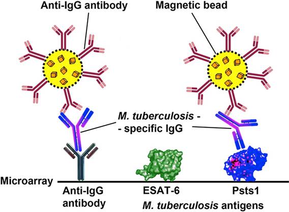
Schematic diagram of a quantitative ultrasensitive immunoassay for immunoglobulins specific to M. tuberculosis antigens
The results are published in Analytica Chimica Acta, impact factor: 4.95.
Shlyapnikov Y.M., Morozov V.N. Titration of trace amounts of immunoglobulins in a microarray-based assay with magnetic labels. Anal.Chim.Acta 2017, 966, 47-53.
A prototype of multi-enzyme sensors and microreactors was fabricated, consisting of a combination of oppositely charged polyelectrolyte layers and enzyme-containing microcapsules. The capabilities of multi-enzyme systems for detecting substances in biological fluids (blood, urine) were investigated. A software was developed and tested for the numerical display of the detected concentrations of two blood metabolites.
(ITEB RAS, Fomkina M. G., Ph.D in Biology)


